

Introduction
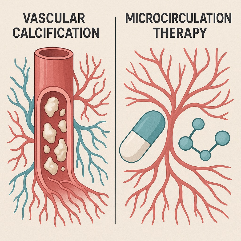
Calciphylaxis — also known as calcific uremic arteriolopathy — represents one of the most enigmatic and devastating vascular complications encountered in nephrology and urology. This rare condition, marked by progressive calcification and thrombosis of dermal and subdermal arterioles, leads to painful necrotic lesions that challenge even the most aggressive therapeutic strategies. While calciphylaxis predominantly afflicts the lower limbs, its occurrence in the penile vasculature is an even rarer and more alarming manifestation. When it does occur, penile calciphylaxis often signals systemic vascular catastrophe and portends a grim prognosis.
The disease typically affects patients with end-stage renal disease (ESRD) undergoing chronic hemodialysis or those who have received renal transplants. It emerges as a paradox of modern medicine: a product of both advanced dialysis survival and the biochemical turmoil that such survival entails — disordered calcium-phosphate metabolism, secondary hyperparathyroidism, and chronic inflammation. Despite decades of case documentation, effective treatment remains elusive.
Amidst this bleak therapeutic landscape, an unconventional pharmacologic candidate has emerged: Tadalafil, a phosphodiesterase type 5 (PDE5) inhibitor better known for its role in erectile dysfunction. While the idea of using a PDE5 inhibitor for a necrotic vascular condition may appear counterintuitive, the mechanistic rationale is elegantly simple — Tadalafil enhances microvascular perfusion by promoting nitric oxide-mediated vasodilation and may thereby counteract ischemia in calcified penile tissue.
A recent case from Chile, reported by Carlos Castro Moya et al. (2019), brings this idea to life. The report describes a man with ESRD and penile calciphylaxis who achieved near-complete remission of ulcerative lesions following daily Tadalafil therapy. This article delves into the underlying science, clinical implications, and broader lessons of this case — bridging vascular pathology, pharmacologic creativity, and clinical pragmatism.
Understanding Calciphylaxis: A Pathology of Vascular Betrayal
A Disease of Paradox
Calciphylaxis is, in essence, a vascular paradox. It arises not from calcium deficiency but from pathologic calcium overload, where the body’s most vital mineral becomes its own worst enemy. Instead of fortifying bone, calcium deposits in small arteries and arterioles, turning pliant microvasculature into rigid conduits incapable of delivering oxygen. The skin and subcutaneous tissues — richly vascularized yet exquisitely dependent on small-artery flow — become prime victims.
Histologically, calciphylaxis is defined by:
- Medial calcification of small arterioles
- Intimal hyperplasia
- Thrombosis and ischemic necrosis of the dermis and subcutis
- Extravascular calcium deposition
Clinically, this manifests as painful, violaceous plaques that progress into necrotic ulcers, often colonized by bacteria, leading to sepsis — the principal cause of death in affected patients. Mortality exceeds 50% within one year, despite intensive wound care and systemic interventions.
Penile Calciphylaxis: An Uncommon Battlefield
Penile involvement is exceptionally rare, with an estimated incidence of around 6% among dialysis patients diagnosed with calciphylaxis. Yet when it occurs, the implications are catastrophic. The penis, with its dense microvascular network and high oxygen demand, becomes particularly vulnerable to arteriolar calcification and ischemia. Patients typically present with painful ulcers, glans necrosis, or dry gangrene, often progressing rapidly despite standard interventions.
The symbolic cruelty of this condition cannot be overstated: in addition to physical agony, it undermines bodily integrity, identity, and dignity — striking at the intersection of survival, sexuality, and self-perception.
Etiopathogenesis: When Mineral Metabolism Turns Malignant
The pathophysiology of calciphylaxis remains incompletely understood, but several interlocking mechanisms have been proposed.
- Disordered Calcium-Phosphate Homeostasis
In ESRD, diminished renal clearance of phosphate and impaired vitamin D synthesis lead to chronic hyperphosphatemia and secondary hyperparathyroidism. Elevated parathyroid hormone (PTH) increases bone resorption, releasing calcium and phosphate into circulation. The resulting calcium-phosphate product precipitates within vascular walls, initiating calcification. - Loss of Endogenous Calcification Inhibitors
The body normally protects against vascular calcification through proteins such as fetuin-A and osteoprotegerin. These inhibitors bind calcium and prevent its deposition in soft tissues. Chronic inflammation in ESRD reduces their synthesis, tipping the balance toward mineral precipitation. - Endothelial Dysfunction and Inflammation
Inflammatory cytokines, particularly interleukin-6 and tumor necrosis factor-alpha, promote endothelial injury and osteogenic transformation of vascular smooth muscle cells. Essentially, arteries begin to behave like bone, depositing calcium along their walls. - Pro-Thrombotic Milieu
Calcified, inflamed vessels become pro-thrombotic. Microvascular thrombosis compounds ischemia, leading to ulceration, necrosis, and secondary infection.
Thus, calciphylaxis represents a perfect storm of metabolic imbalance, inflammatory injury, and vascular rigidity — a process where the blood vessels themselves become ossified monuments to systemic disease.
Case Synopsis: When Tadalafil Defied Expectations
The Chilean case offers an illustrative clinical narrative. The patient, a 57-year-old man with poorly controlled type 2 diabetes mellitus and hypertension, developed ESRD secondary to diabetic nephropathy. He was receiving regular hemodialysis when he presented with three painful, non-healing ulcers on the glans penis. These lesions, covered by yellow-white necrotic tissue, had persisted for two months and resisted conventional measures.
Radiographic imaging revealed extensive calcification of penile vessels, confirming penile calciphylaxis. Laboratory evaluation showed a serum creatinine of 5.7 mg/dL and a parathormone level markedly elevated at 351 pg/mL — consistent with secondary hyperparathyroidism, though calcium-phosphate levels remained within normal limits.
Given the ischemic nature of his condition, the medical team initiated Tadalafil 5 mg daily, targeting microvascular perfusion rather than mineral metabolism. Remarkably, after six months, the ulcers demonstrated near-total healing, and the patient reported complete resolution of pain. Over 18 months of follow-up, with continued hemodialysis and daily Tadalafil, no recurrence was observed.
This outcome stands as a rare victory in a disease where most interventions yield only modest reprieve.
Mechanistic Rationale: How PDE5 Inhibition May Rescue Ischemic Tissue
Beyond Erectile Function
Phosphodiesterase type 5 (PDE5) inhibitors — including sildenafil, vardenafil, and tadalafil — are best known for treating erectile dysfunction. However, their therapeutic value extends far beyond penile rigidity. PDE5 inhibitors act by preserving cyclic guanosine monophosphate (cGMP) levels, thereby amplifying nitric oxide (NO)–mediated vasodilation. The result is improved endothelial function and blood flow across multiple vascular beds.
Microvascular Implications in Calciphylaxis
In calciphylaxis, the chief pathology is microvascular obstruction and ischemia. By promoting smooth muscle relaxation and enhancing perfusion, Tadalafil may:
- Increase oxygen delivery to ischemic tissues
- Reduce vascular resistance and endothelial stress
- Counteract localized thrombosis through mild antiplatelet effects
- Support angiogenesis and capillary remodeling
Moreover, PDE5 inhibitors have demonstrated anti-inflammatory and anti-fibrotic effects in preclinical models, potentially mitigating endothelial injury and vascular stiffening.
Tadalafil’s long half-life (17–36 hours) allows for stable plasma concentrations, providing continuous vasodilatory support — an advantage over shorter-acting PDE5 inhibitors. Its established safety profile in cardiovascular and renal-compromised patients further supports its off-label exploration in this context.
Reframing the Mechanism: A Microcirculatory Perspective
From a physiological viewpoint, Tadalafil does not “cure” calciphylaxis — it buys time. By enhancing perfusion, it may prevent ischemic escalation, allowing existing ulcers to heal and new lesions to be forestalled. The therapy’s success likely hinges on early intervention, before irreversible necrosis sets in. Once extensive gangrene develops, surgical debridement or partial penectomy remains the only recourse.
Diagnosis: When Radiographs Tell the Tale
Diagnosing calciphylaxis demands a blend of clinical suspicion and confirmatory imaging. The hallmark is painful, violaceous skin lesions that evolve into ulcers. In penile disease, these ulcers typically localize to the glans or distal shaft.
The most sensitive diagnostic tool is plain radiography using mammographic technique, capable of revealing fine vascular calcifications. In the Chilean case, pelvic X-ray provided clear visualization of calcified penile arteries — a finding that, though pathognomonic, often arrives late in the disease course.
Laboratory markers are nonspecific. Calcium and phosphate levels may paradoxically remain normal, as vascular deposition depletes circulating calcium. Thus, normal lab values do not exclude calciphylaxis. Histological confirmation — while definitive — carries risk, as biopsy can precipitate infection or worsen necrosis, particularly in penile tissue.
Differential diagnoses include:
- Vasculitic syndromes
- Cryoglobulinemia
- Atheroembolism
- Coumarin necrosis
- Necrotizing fasciitis
Yet none of these conditions exhibit the arteriolar calcification that defines calciphylaxis.
Current Therapeutic Landscape: A Grim Reality
Calciphylaxis management remains largely empirical and supportive. Standard treatments aim to correct metabolic derangements and control pain, but outcomes are disheartening. Therapeutic modalities include:
- Phosphate binders (e.g., sevelamer, lanthanum carbonate) to reduce phosphate burden
- Calcimimetics (e.g., cinacalcet) to suppress PTH secretion
- Sodium thiosulfate as a chelating agent for vascular calcium deposits
- Bisphosphonates, inhibiting osteoclastic activity and vascular calcification
- Parathyroidectomy, reserved for refractory secondary hyperparathyroidism
Despite these efforts, the one-year survival rate for calciphylaxis remains below 50%, largely due to sepsis from infected ulcers. Pain control and wound care, though palliative, dominate clinical management.
Against this background, the Tadalafil case represents an innovative departure — not targeting calcium metabolism, but improving perfusion directly.
Discussion: Lessons from an Unlikely Success
Why Tadalafil Worked — and Why It Might Not Always
The Chilean case underscores a fundamental truth in vascular medicine: sometimes, flow matters more than chemistry. By improving penile microcirculation, Tadalafil likely tipped the physiological balance away from ischemia and toward repair. Yet this success is not universally guaranteed. The therapy may only be effective in early, non-gangrenous stages, where viable tissue remains.
Moreover, the rarity of penile calciphylaxis means clinical trials are improbable. Each case thus becomes a mini-experiment in applied physiology, guided by the clinician’s judgment and the patient’s resilience.
The Broader Implications for Microvascular Medicine
This case also invites broader reflection. PDE5 inhibitors are already used in conditions like pulmonary arterial hypertension, Raynaud’s phenomenon, and chronic wound ischemia, where microvascular flow is compromised. Extending their use to calciphylaxis is not a conceptual leap, but rather a logical progression — one that aligns pharmacologic principles with microvascular pathology.
In this sense, Tadalafil’s role in calciphylaxis is not an outlier, but a natural extrapolation of its vascular effects.
Ethics and Innovation in End-Stage Disease
Off-label use of established drugs often raises ethical questions. Yet in terminal conditions with no effective therapies, innovation becomes an ethical imperative. The use of Tadalafil in this context exemplifies rational compassion — leveraging existing pharmacology to restore dignity, comfort, and perhaps life itself.
Prognosis: Between Hope and Realism
Even with optimal management, calciphylaxis remains a lethal disease. Survival depends on early recognition, aggressive infection control, and careful wound management. The prognosis worsens with delayed diagnosis or extensive necrosis.
In the reported case, survival beyond 18 months with sustained tissue integrity is noteworthy. Whether Tadalafil altered disease trajectory or merely optimized local perfusion remains uncertain. Nonetheless, the absence of ulcer recurrence over prolonged follow-up strongly suggests a durable therapeutic benefit.
Conclusion
Penile calciphylaxis stands as a grim reminder of how metabolic chaos can manifest as vascular catastrophe. In this context, the innovative use of Tadalafil represents more than pharmacologic curiosity — it symbolizes the clinician’s enduring search for light in the vascular darkness of ESRD.
By enhancing microvascular perfusion, Tadalafil may prevent tissue loss, relieve pain, and restore function, particularly when initiated early. While not curative, it offers a pragmatic means of extending survival and improving quality of life in a condition where conventional therapies falter.
Ultimately, this case challenges the boundaries of urological medicine and underscores a universal lesson: sometimes, healing begins not by fighting the disease itself, but by restoring what the disease has taken away — circulation, oxygen, and hope.
FAQ
1. What exactly is penile calciphylaxis?
It is a rare form of vascular calcification affecting small arteries in the penis, typically in patients with end-stage renal disease. The resulting ischemia leads to painful ulceration and tissue necrosis.
2. How can Tadalafil help in such a condition?
Tadalafil improves blood flow by enhancing nitric oxide–mediated vasodilation. In early calciphylaxis, this may counteract ischemia and promote healing of ulcerated tissue, though it does not reverse existing calcifications.
3. Is Tadalafil now a standard treatment for calciphylaxis?
No. Its use remains experimental and based on isolated case reports. However, given its favorable safety profile and plausible mechanism, it represents a promising adjunct in carefully selected patients.
