

Introduction: Oral Bioavailability Is Decided in a Fluid You Rarely See
Oral administration remains the most practical and patient-friendly route for systemic therapy, but it comes with a stubborn rule: a drug must dissolve before it can be absorbed. For BCS class II compounds—high permeability, low solubility—this rule becomes the entire game. We can engineer elegant tablets, coat them beautifully, and write confident labels, yet the drug’s performance still hinges on what happens in the intestinal lumen, in a fluid whose composition changes from person to person and hour to hour.
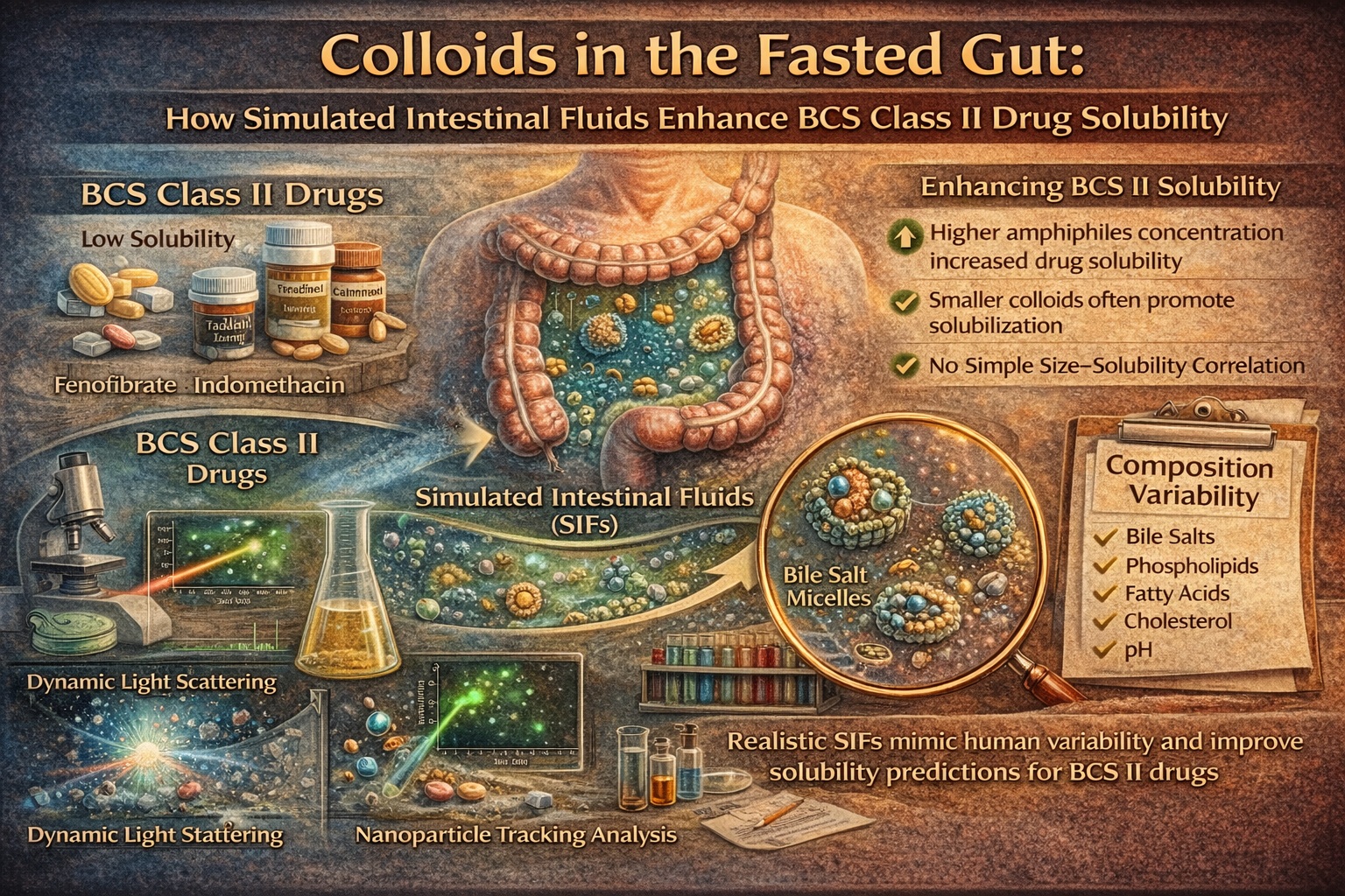
That intestinal fluid is not a simple buffer. It contains bile salts, phospholipids, fatty acids, cholesterol, salts, and variable pH, and these components self-assemble into colloidal structures—mixed micelles, vesicles, and aggregates—that can dramatically raise the apparent solubility of lipophilic drugs. In other words, the gut creates its own solubilizing system, but the “settings” are different in every individual and prandial state.
A common limitation in formulation science is the reliance on a single fasted-state simulated intestinal fluid (FaSSIF) as a stand-in for the entire population. It is convenient, standardized, and reproducible—three virtues that often hide a fourth characteristic: it may not represent the variability that actually controls solubilization in vivo.
A 2024 experimental study tackled this problem directly by building a suite of fasted-state simulated intestinal fluids (SIFs) reflecting variability seen in human intestinal fluid, then testing solubility of eight poorly soluble drugs (acidic, basic, and neutral) while characterizing colloidal structures using both dynamic light scattering (DLS) and nanoparticle tracking analysis (NTA). The results are a useful correction to simplistic assumptions: solubilization often increases with amphiphile concentration, colloidal size changes dramatically with composition, and yet the relationship between “smaller particles” and “higher solubility” is not universally straightforward.
Why “One FaSSIF” Is a Dangerous Comfort: Composition Variability Drives Outcomes
Fasted human intestinal fluid is variable in ways that matter pharmacokinetically. pH can shift across a meaningful range, and amphiphile concentrations (bile salts, phospholipids, and fatty acids) can vary substantially. These variations alter the total amphiphile concentration (TAC) and, by extension, the number and nature of colloidal solubilizing structures available.
The study built multiple SIF recipes spanning low to high pH × TAC conditions, including a “minimum” point with very low amphiphile content and a “maximum” point with very high amphiphile content (so high that it begins to resemble fed-state behavior). This is a pragmatic approach: rather than pretending the gut is constant, it tries to emulate how different a “fasted gut” can look across individuals.
Why does this matter clinically? Because the same BCS class II drug can behave very differently across patients even when permeability is high. A formulation that performs well in a single biorelevant medium may underperform in a patient whose intestinal amphiphile concentration is lower, whose pH differs, or whose colloidal structures are less favorable for drug incorporation.
The study’s central message is that variability is not noise. It is a mechanistic input. If solubility is the limiting step, and solubility depends on colloidal structures, then a realistic in vitro model must allow those structures to change the way they do in humans.
Colloidal Structures: Micelles, Vesicles, and the Chemistry of “Helping a Drug Dissolve”
Bile salts and phospholipids do not merely float around politely. As concentrations rise toward and beyond the critical micelle concentration (CMC), bile salts self-assemble into aggregates and form mixed systems with phospholipids and other amphiphiles. Below the CMC, bile salts can insert into phospholipid vesicles without destroying them; above it, they promote mixed micelles and other morphologies capable of solubilizing lipophilic compounds.
Cholesterol adds further complexity, influencing membrane packing and colloid stability. The classic versions of FaSSIF have historically omitted cholesterol, which may be reasonable for standardization but is less faithful to biology. In a real intestine, cholesterol is part of the amphiphile landscape and can shift colloidal structure behavior.
From a drug’s perspective, these colloids offer “hydrophobic real estate.” Lipophilic molecules can partition into hydrophobic domains of micelles or bilayer structures, raising apparent solubility. For weak acids or bases, ionization state adds an additional layer: ionized species may associate differently (often nearer the hydrophilic interface), potentially affecting colloid size and interactions.
What is striking in this work is not the existence of colloids—it is how drastically their measured sizes shift with changing composition, and how drug presence itself can modify the colloidal population. The gut is not a passive container; it is a dynamic self-assembling solvent system that responds to what you put into it.
What the Study Found: Solubility Rises with Amphiphiles, but Particle Size Tells a Complicated Story
Across the suite of fasted-state SIFs, solubility generally increased as TAC increased, with a clear pattern: acidic drugs were more soluble than neutral or basic drugs in the media evaluated. This is consistent with the role of pH-driven ionization for weak acids, especially as pH rises above pKa, and also reflects the different ways neutral and basic drugs depend on amphiphile-driven solubilization rather than purely on ionization.
A key methodological strength is that the authors did not only measure solubility; they measured colloidal size distributions in the presence and absence of each drug using DLS and (for the first time in this context) NTA. DLS revealed that as amphiphile concentration increased, the hydrodynamic diameters tended to decrease. The fluids were also notably polydisperse, unlike the more monodisperse behavior often observed with standard FaSSIF, reminding us that “clean” size distributions may reflect the simplicity of the model rather than the reality of the intestine.
The magnitude of size change can be dramatic. For example, in the presence of felodipine, structures measured in minimum TAC media were around 170 nm, falling to roughly 5 nm in maximum TAC conditions. Standard FaSSIF (used as a comparator) produced intermediate behavior. This wide range highlights that colloids are not a single entity—they are an ensemble whose dominant species changes with composition.
Yet the most important scientific honesty in the study is this: there was no simple correlation between solubility and colloidal size. Although high solubility often coincided with smaller measured colloids in DLS, the relationship was not universal across drug classes and compositions. Some drugs showed unusual behavior—carvedilol, for instance, produced very large apparent structures in certain media and displayed solubility behavior that did not follow the naïve “pH above pKa means lower solubility” expectation, indicating strong amphiphile-driven solubilization effects that can overpower simple ionization logic.
In practical terms, “smaller micelles = more solubilization” is a tempting slogan, but slogans do not dissolve drugs. Colloidal morphology, polydispersity, drug–colloid affinity, and phase behavior can each dominate depending on the system.
DLS vs NTA: Two Instruments, Two Realities, One Lesson About Measurement Bias
Dynamic light scattering is widely used for particle sizing in colloidal systems, but it has a known weakness: it is highly sensitive to larger particles. Even a small population of aggregates can dominate the scattering signal and inflate apparent size. In complex, polydisperse intestinal media—where dust, aggregates, and heterogeneous structures exist—this matters.
NTA, on the other hand, tracks particle motion under laser illumination and can detect small, weakly scattering particles among larger ones. The study used NTA to characterize SIF colloids and compared results with DLS, finding significant differences between measurements in every drug/media combination tested.
A clinically relevant nuance emerges: NTA requires dilution to achieve measurable particle concentrations, but dilution itself can perturb colloids, particularly those near the CMC or formed at high amphiphile concentrations. This means NTA can underestimate or reshape the very structures we wish to measure—especially in high-TAC systems where dilution may push the sample below aggregation thresholds.
The authors observed that the largest discrepancies between DLS and NTA appeared in higher-TAC media points. DLS detected very small sizes (around 5–6 nm) in maximum conditions, while NTA frequently measured much larger modal sizes, likely influenced by dilution-driven structural changes.
The practical takeaway is not that one technique is “right” and the other “wrong.” The takeaway is that measurement defines reality in colloidal science. When linking colloidal properties to solubility, the method—and its biases—must be treated as part of the model. Otherwise, we risk explaining pharmacokinetics with artifacts.
Implications for Formulation and Translational Prediction: From “Average Media” to Patient-Reflective Testing
For formulation scientists, these findings support a shift from single-point testing toward variability-aware biorelevant testing. If a BCS II drug’s solubility increases with TAC, and TAC varies across individuals, then dissolution performance may vary in a way that standard media cannot reveal. That is not a theoretical concern; it is the kind of variability that drives “works in trials, inconsistent in practice” outcomes.
This is especially relevant when developing formulations that rely on intestinal colloids for solubilization rather than generating their own solubilizing environment (e.g., lipid-based formulations, amorphous dispersions with bile interactions, or drug products that depend on micellar uptake). A formulation may look excellent in standard FaSSIF, yet fail in low-TAC conditions that occur in certain fasted individuals.
The study also reminds clinicians—indirectly but importantly—that food effects are not merely about “fed vs fasted.” Even within the fasted state, physiologic variability can change solubilization capacity. If a drug has a narrow therapeutic window or requires consistent exposure, understanding its dependence on intestinal amphiphiles becomes more than an academic exercise.
If you want to operationalize the study’s lessons, the most reasonable framework is to treat SIF composition and colloidal characterization as part of a decision pipeline rather than a single confirmatory experiment:
- Use a suite of fasted-state SIFs spanning realistic pH × TAC variability to map solubility ranges.
- Characterize colloidal structures with at least two complementary techniques, acknowledging their biases.
- Interpret solubility trends by drug class (acidic/basic/neutral), but avoid assuming a universal size–solubility rule.
- Use outliers (e.g., drugs showing unexpected solubility increases at high pH due to amphiphiles) as signals for deeper mechanistic study rather than “bad data.”
- Design formulations to be robust across low-solubilization conditions, not merely optimized for average media.
A mild irony fits here: we often design “patient-centered” medicines, but test them in intestinal fluids that represent a patient who does not exist—an average composite with an unusually stable gut. Variability-aware testing is less glamorous than novel excipients, but far more likely to prevent unpleasant surprises.
Conclusion: The Gut’s Colloids Are Not Background—They Are a Biopharmaceutic Determinant
This work reinforces a central reality of oral pharmacotherapy: for BCS class II drugs, intestinal colloids are not incidental. They are one of the primary determinants of how much drug becomes available for absorption. The study shows that fasted-state simulated intestinal fluids can be engineered to reflect real human variability, and that this variability meaningfully impacts drug solubility.
It also demonstrates that colloidal structures shift substantially with composition and drug presence, and that different measurement methods can yield very different size interpretations—especially in polydisperse, high-amphiphile systems. The relationship between colloid size and solubilization is real, but not simplistic; it is mediated by structure type, composition, polydispersity, and drug–colloid interactions.
For scientists developing oral medicines, the message is practical: build predictive models that tolerate biological variability, validate with multiple particle characterization techniques, and treat “no simple correlation” not as disappointment, but as accurate reporting of a complex physiological system.
In short, the intestine is not a beaker, and its colloids are not optional. If we want oral therapies to behave predictably in patients, our in vitro systems must be brave enough to look messy.
FAQ
1. Why are BCS class II drugs so sensitive to intestinal fluid composition?
Because absorption is usually limited by dissolution and solubilization. Changes in bile salts, phospholipids, fatty acids, cholesterol, and pH alter the gut’s colloidal structures that help dissolve lipophilic drugs.
2. Does smaller colloidal particle size always mean higher drug solubility?
Not necessarily. While higher amphiphile concentrations can produce smaller structures and often increase solubility, the study shows no universal size–solubility rule. Morphology, polydispersity, and drug–colloid affinity can dominate.
3. Why do DLS and NTA sometimes disagree on particle size in SIF?
DLS is highly influenced by larger scatterers and can overrepresent aggregates, while NTA requires dilution that can disrupt colloids—especially near the CMC or in high-amphiphile media—leading to different apparent size distributions.
