

Introduction
Temporal lobe epilepsy (TLE) remains the most common form of focal epilepsy in adults, accounting for a significant proportion of drug-resistant cases. For decades, the clinical narrative has been dominated by seizures refractory to medical therapy, variable neuropsychological impairment, and the tantalizing promise of surgical cure. The challenge, however, lies in identifying the precise structural and functional abnormalities within the temporal lobe that drive epileptogenesis.
Standard MRI protocols often reveal hippocampal sclerosis, cortical malformations, or neoplastic lesions, all of which guide surgical decisions. Yet in a considerable number of patients, conventional MRI appears “normal,” leaving clinicians in a diagnostic quandary. For these individuals, the risk of misclassification, delayed treatment, or unsuccessful surgery looms large. The urgency of innovation is obvious: without accurate localization, surgical outcomes falter and patient morbidity rises.
Recent advances in MRI technology—ranging from high-field imaging to sophisticated post-processing algorithms—offer a new dimension to the evaluation of TLE. By probing microstructural, functional, and connectivity-related features of the brain, modern MRI transcends the limits of visual inspection, turning the invisible into the clinically actionable. The emerging message is clear: advanced MRI is no longer a research luxury but a clinical necessity in temporal lobe epilepsy.
The Clinical Burden of Temporal Lobe Epilepsy
Temporal lobe epilepsy is not merely an episodic neurological disturbance. It is a chronic disease with pervasive implications for cognition, behavior, and quality of life. The recurrent seizures often resist pharmacological control, leading to the classification of drug-resistant epilepsy in nearly one-third of patients. These individuals endure persistent disability, increased risk of injury, and heightened mortality compared with the general population.
Cognitively, TLE exerts a profound toll. The hippocampus, central to memory processing, is frequently implicated, and repeated seizures exacerbate memory decline. Language deficits, emotional dysregulation, and executive dysfunction are common companions, reflecting the intricate network connections of the temporal lobe. Thus, the disease trajectory extends beyond seizures to encompass a spectrum of neurological deterioration.
Surgically, anterior temporal lobectomy and selective amygdalohippocampectomy remain the mainstays of therapy, offering seizure freedom in many cases. Yet surgical success hinges on precise localization of the epileptogenic zone. Patients with clear MRI abnormalities, such as hippocampal sclerosis, generally fare better postoperatively. By contrast, those with inconclusive imaging results often confront disappointing outcomes, underscoring the clinical imperative for advanced diagnostic methods.
Conventional MRI: Strengths and Shortcomings
For years, conventional MRI has been the workhorse of epilepsy evaluation. T1-weighted, T2-weighted, and FLAIR sequences reveal gross structural anomalies, providing critical information for pre-surgical planning. The sensitivity of MRI for hippocampal sclerosis, for instance, is well established, with findings such as reduced hippocampal volume and increased T2 signal serving as classic hallmarks.
However, the limitations of routine MRI are equally well recognized. Up to 30% of TLE patients exhibit “MRI-negative” findings, despite clear electroclinical evidence of temporal lobe origin. These patients often undergo invasive intracranial EEG to compensate for the lack of structural guidance—a procedure that, while valuable, carries inherent risks. Furthermore, subtle cortical dysplasias, microstructural abnormalities, and network-level dysfunctions frequently escape the resolution of standard MRI protocols.
The consequence is diagnostic uncertainty. When MRI fails, clinicians face the dilemma of whether to proceed with invasive testing, adopt a conservative therapeutic stance, or risk surgery with incomplete information. Each option carries potential harm, emphasizing the pressing need for tools that extend MRI beyond the visible.
Advanced MRI Techniques: A Paradigm Shift
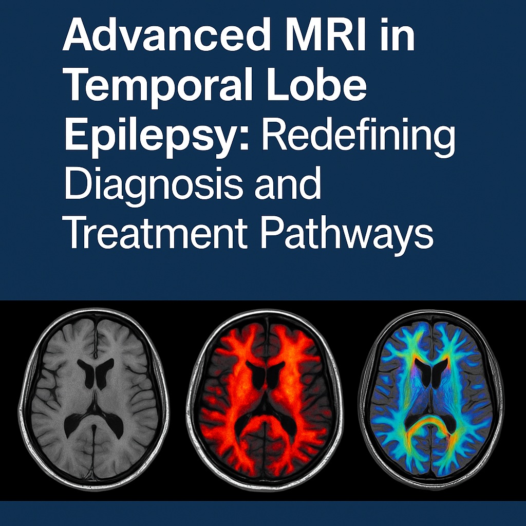
Recent innovations in MRI are transforming the diagnostic landscape of temporal lobe epilepsy. Unlike conventional imaging, which captures macroscopic anatomy, these methods probe tissue microstructure, functional dynamics, and connectivity.
Diffusion tensor imaging (DTI) assesses the integrity of white matter tracts by measuring water diffusion along axonal pathways. In TLE, abnormalities in the fornix, cingulum, and uncinate fasciculus reveal network-level disruptions that may underlie both seizures and cognitive deficits. These findings extend the diagnostic gaze beyond the hippocampus, illustrating epilepsy as a disorder of networks rather than isolated lesions.
Functional MRI (fMRI) further enriches this perspective by mapping brain activity through blood oxygenation level-dependent signals. Task-based fMRI aids in language and memory lateralization, guiding surgical planning to minimize postoperative deficits. Resting-state fMRI uncovers aberrant connectivity patterns, often concordant with the epileptogenic zone, and holds promise as a noninvasive adjunct to EEG.
Meanwhile, high-field MRI at 7 Tesla enhances spatial resolution, unveiling cortical dysplasias and subtle hippocampal anomalies previously undetectable. Post-processing techniques, such as voxel-based morphometry and automated volumetric analyses, enable quantitative assessment of gray matter changes, reducing reliance on subjective visual interpretation. Together, these innovations form a multimodal arsenal for deciphering the enigmatic brain of TLE patients.
Correlation With Clinical Outcomes
The true test of any diagnostic innovation lies in its clinical utility. Advanced MRI has demonstrated clear value in predicting surgical outcomes for TLE. Patients with abnormalities detected by DTI or volumetric analyses, even in the absence of conventional MRI findings, often achieve seizure freedom comparable to those with visible lesions. This underscores the sensitivity of advanced imaging in identifying epileptogenic substrates.
Cognitive outcomes also correlate with MRI findings. For instance, preoperative fMRI can predict the risk of postoperative memory decline by assessing hippocampal activation patterns. Similarly, diffusion abnormalities in memory-related tracts foreshadow cognitive deficits, allowing clinicians to balance seizure control with preservation of function.
Perhaps most compelling is the synergy between MRI and other modalities. When combined with EEG, PET, or magnetoencephalography, advanced MRI enhances diagnostic confidence, reduces reliance on invasive testing, and streamlines surgical decision-making. The convergence of multimodal data is increasingly recognized as the optimal strategy for maximizing both seizure and cognitive outcomes.
Challenges and Limitations
Despite its promise, advanced MRI is not without obstacles. Technical variability across centers limits standardization, making it difficult to compare results or establish universal thresholds. The high cost and limited availability of 7 Tesla MRI restrict access to specialized centers, raising concerns about equity in care. Furthermore, advanced image analysis demands expertise in neuroimaging and computational methods, skills not universally available in clinical practice.
Interpretation also remains a challenge. Not every abnormality detected by advanced MRI is epileptogenic, and false positives can mislead surgical planning. Distinguishing causative lesions from incidental findings requires careful integration of clinical, electrophysiological, and imaging data.
Finally, ethical considerations loom large. The proliferation of imaging biomarkers raises questions about incidental discoveries—findings unrelated to epilepsy but potentially clinically significant. Balancing patient autonomy with the responsibility to disclose uncertain results is a nuanced and ongoing debate in neuroimaging ethics.
Future Directions
The trajectory of advanced MRI in temporal lobe epilepsy points toward increasing sophistication and integration. Machine learning algorithms are already being deployed to analyze high-dimensional imaging data, offering automated classification of patients into surgical candidates or non-candidates. These models promise to reduce inter-observer variability and accelerate diagnostic workflows.
In parallel, hybrid imaging approaches combining MRI with PET tracers are under investigation, aiming to link structural and metabolic insights into a unified diagnostic picture. Moreover, longitudinal MRI studies are beginning to unravel the progressive nature of network alterations in TLE, shedding light on the dynamics of epileptogenesis and treatment response.
Ultimately, the future lies in personalization. By tailoring diagnostic pathways to individual imaging profiles, clinicians can move beyond “one-size-fits-all” strategies to precision medicine in epilepsy. This vision entails not only better seizure control but also preservation of cognitive function, improved quality of life, and reduced treatment delays.
Conclusion
Temporal lobe epilepsy challenges clinicians with its heterogeneity, its cognitive burden, and its stubborn resistance to medical therapy. Yet it also offers a unique therapeutic opportunity: surgery can be curative if the epileptogenic zone is identified with confidence. Advanced MRI has emerged as a pivotal tool in this quest, revealing structural and functional abnormalities invisible to conventional imaging and guiding surgical strategies with unprecedented precision.
The message is both hopeful and urgent. Hopeful, because modern imaging can unlock seizure freedom for patients once deemed inoperable. Urgent, because failure to adopt these technologies risks perpetuating diagnostic uncertainty and therapeutic inertia. In the landscape of temporal lobe epilepsy, advanced MRI is not an optional upgrade but an essential evolution.
FAQ
1. Why do some patients with temporal lobe epilepsy have “normal” MRI scans?
Because conventional MRI often lacks the resolution to detect subtle cortical dysplasias, microstructural abnormalities, or network dysfunctions. Advanced techniques such as DTI and high-field imaging can reveal these hidden lesions.
2. How does advanced MRI improve surgical outcomes?
By providing precise localization of the epileptogenic zone and predicting cognitive risks, advanced MRI allows surgeons to tailor resections that maximize seizure freedom while minimizing functional deficits.
3. Is advanced MRI widely available for epilepsy patients?
Access remains limited to specialized centers, particularly for high-field MRI. However, diffusion imaging, volumetric analysis, and fMRI are increasingly incorporated into routine epilepsy evaluations worldwide.
